Digital Trichoscopy & Scalp Analysers: A 2026 Clinic Guide
A practical guide to adding doctor-led scalp imaging: recent global and Indian research, equipment checks, repeatable records, privacy and realistic service costs.
Research checked: 26 October 2026. For Indian dermatologists, aesthetic doctors and clinic owners.
A scalp analyser can make hair consultations easier to document, but a useful hair service requires more than a magnified picture. Doctors need a clinical history, an examination and a reason for choosing each investigation. The equipment should support those decisions and help patients understand their follow-up plan.
For a clinic planning its next service, digital scalp imaging offers a practical assessment option to evaluate before purchasing additional treatment equipment. The aim is a repeatable baseline and a doctor-led review, rather than selling every patient the same hair package. This guide looks at current research, buying questions and a proposed service model for Indian practices.
What is new in scalp imaging research?
An international research preprint submitted on 1 October 2026 describes an AI framework for analysing low-magnification scalp images. Its components include follicular-unit detection, visible hair-shaft counting and width estimation. The authors explicitly position the system as assistance for dermatologist interpretation. An arXiv preprint is preliminary research, not evidence that every commercial AI scalp analyser has equivalent performance or authorization. Source: Raslan and colleagues [1].
There is also recent Indian research using trichoscopy to measure treatment outcomes. A study published on 25 September 2026 from investigators in Ujjain compared platelet-rich plasma and injectable platelet-rich fibrin in 35 men with androgenetic alopecia. It used trichoscopic hair-density assessment. Fixed allocation to scalp sides, no documented formal blinding and short follow-up limit interpretation. It does not establish that either treatment suits every patient or validate a particular imaging machine. Source: Central India Journal of Medical Research [2].
These examples show interest in measurable scalp assessment. For a buyer, the useful question is whether a specific system delivers reliable information for the clinic's intended use. A research result should not become an unsupported promise on a consultation report.
Start with diagnosis, then choose the imaging
The American Academy of Dermatology explains that finding the cause is central to hair-loss treatment. Assessment includes questions, examination and, when clinically indicated, additional investigations such as blood tests or a scalp biopsy. Imaging cannot replace this wider process. Source: AAD hair-loss guidance [3].
Trichoscopy is dermoscopic examination of the scalp and hair. A device marketed as a scalp analyser may offer magnification, photographs or automated scores, but those functions are not interchangeable. Ask the supplier to describe what is observed, what is measured and what is inferred by software.
A clinician should decide which patients need an imaging record, how findings fit the examination and when further assessment is necessary. Avoid describing a generic image score as proof of a nutritional deficiency, a hormonal diagnosis or the guaranteed need for a procedure.
A practical service to add to the clinic
Consider a structured hair-and-scalp consultation with documented imaging where appropriate. Before the appointment, explain what is included, any fee and who interprets the report. During the visit, the doctor can review the history, examine the patient and decide whether imaging adds useful information.
A baseline record can include agreed scalp sites, consistent overview photographs and selected magnified images. The patient should receive an understandable explanation of the findings, the clinical plan and the purpose of follow-up. A normal-looking image should not override symptoms or clinical concerns.
At subsequent visits, compare like with like. Changes in hairstyle, lighting, magnification or the photographed site can make images look different without proving a biological change. Do not attribute improvement to a treatment solely because two photographs appear different.
Five buying checks for a digital scalp analyser
1. Image capture and repeatability
Request a demonstration using a written capture method. Evaluate focus, illumination, magnification, positioning and the ability to return to the same site. Ask how image scale is calibrated if the software reports measurements. Keep original images available for clinical review rather than relying entirely on an attractive summary PDF.
2. Evidence for automated measurements
Ask for validation of the exact model and software version. Which outputs were compared with expert assessment? What happens with blurred images, hair products or difficult capture conditions? Request information relevant to the clinic's patient population. Research on another algorithm does not automatically apply to the proposed machine.
3. Indian documentation and intended use
CDSCO describes medical devices in terms of their intended purposes, including diagnostic uses, and includes software in its definition. Review the exact system's intended use and applicable Indian documentation with the supplier. Seek qualified regulatory advice where classification or authorization is unclear. Source: CDSCO [4].
Do not assume that the words AI, professional or imported establish medical validity. Match the manufacturer's identity, model and supplied configuration to the documents. Avoid advertising an approval or certification until its relevance to that exact product has been verified.
4. Records, privacy and portability
Confirm where patient photographs are stored, who can access them and how they are exported or deleted. Ask whether processing requires a cloud service and whether images may be reused for software development. Establish appropriate consent and access controls before routine use. Obtain separate permission for any marketing use of patient photographs.
5. Training, maintenance and support
Separate training in operating the camera from competence in clinical interpretation. Establish who captures images, who checks quality and which doctor reviews findings. Obtain written details of warranty, software renewals, maintenance and service availability. Ask whether existing records remain accessible if the subscription ends or the device needs repair.
Plan operating costs without assuming demand
Request quotations that include the complete setup: equipment, required computer, software, installation, training and recurring support. Add staff capture time, clinician review time, secure storage, cleaning requirements and any consumables. A low initial price may be less attractive if routine exports or essential software features cost extra.
Decide whether imaging is part of an existing consultation or a separately priced assessment. Estimate completed appointments using the clinic's actual capacity and observed demand. Compare collected revenue with the additional operating costs, and test a lower-volume scenario. There is no basis for promising a fixed payback period or guaranteed additional income.
Use a small pilot to assess usefulness
Begin with a defined appointment type and capture protocol. Track image retakes, consultation duration, record completeness, export reliability and patient understanding. Review whether follow-up images are comparable and whether the service improves the doctor's documentation. Procedure sales alone are not a sufficient measure of clinical value.
Beauty Enrich lists scalp-analysis equipment, including the T8 AI Intelligent Scalp & Hair Analyzer [5]. Use the product page as a starting point for a model-specific demonstration and documentation request. This article does not independently verify its diagnostic accuracy or regulatory status. Our AI skin analysis guide [6] also explains why software outputs and clinical diagnosis must be considered separately.
Frequently asked questions
Can a scalp analyser diagnose every cause of hair loss?
A magnified image or automated report does not replace a doctor's history and examination. Additional investigations may be appropriate depending on the clinical findings. The system should be used within its documented intended purpose.
Is digital trichoscopy a hair-regrowth treatment?
It is an assessment and documentation approach. Buying imaging equipment does not itself provide a treatment or establish that a patient needs one. The doctor determines the care plan.
Should every clinic buy an AI system?
Compare the proposed system with simpler dermoscopic imaging and photography options. Choose according to clinical needs, evidence, record quality, staff competence and ongoing costs. An AI label alone is not a purchasing reason.
How should results be explained to patients?
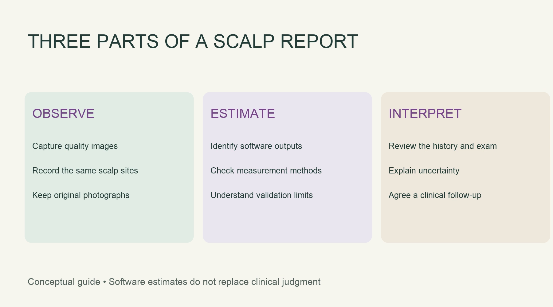
Separate observed features, software estimates and the doctor's interpretation. Explain uncertainties and what will be reviewed at follow-up. Avoid using a numerical score to pressure a patient into a treatment package.
Sources and related reading
[1] Raslan et al. AI-based scalp assessment framework. arXiv preprint submitted 1 October 2026; preliminary research.
https://arxiv.org/abs/2610.02421
[2] Vijayvargiya et al. Prospective split-scalp PRP and PRF study, published 25 September 2026.
https://cijmr.com/index.php/cijmr/article/view/485
[3] American Academy of Dermatology: Hair loss diagnosis and treatment.
https://www.aad.org/public/diseases/hair-loss/treatment/diagnosis-treat
[4] CDSCO: Medical Device and Diagnostics information.
https://www.cdsco.gov.in/opencms/opencms/en/Medical-Device-Diagnostics/Medical-Device-Diagnostics/
[5] Beauty Enrich: T8 AI Intelligent Scalp & Hair Analyzer.
https://www.beautyenrich.com/products/t8-ai-intelligent-scalp-hair-analyzer
[6] Beauty Enrich: AI Skin Analysis Machines: A 2026 Guide for Indian Clinics.
https://www.beautyenrich.com/blogs/ai-skin-analysis-machines-indian-clinics-2026
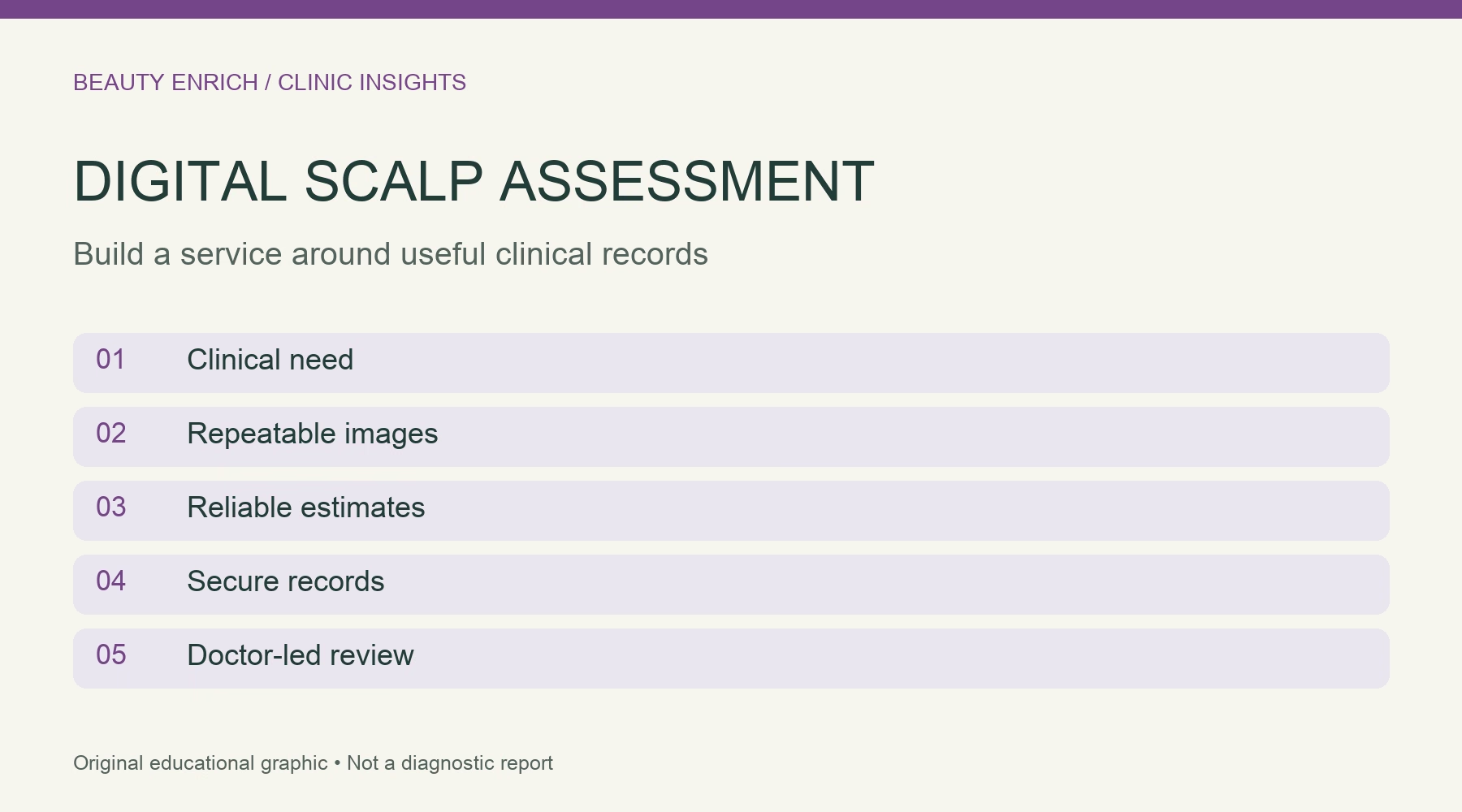
Image captions: The cover maps five considerations for a digital scalp assessment service. The supporting diagram distinguishes observed images, software estimates and clinical interpretation. Both are original educational graphics, not clinical images, diagnostic evidence or validated care protocols.